 Fluorescence ratio measurement
Complete
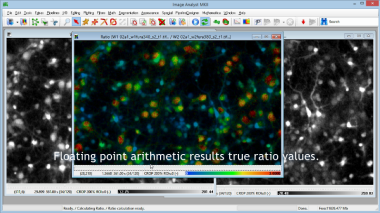
analysis of a fura-2 calcium recording in rat cortical neurons.
Key features:
- Image registration to fix movement of the specimen during pipetting
- Automatic background subtraction
- Automatic single-cell ROI drawing
- True ratio image and ratio calculation from ROI means
- Ratio values are directly transferred to Excel
Run time: 4 min 00 s.
 H2O2 assay using normalized DCFDA rates
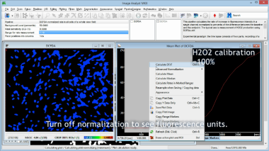
Complete analysis of intracellular H2O2 levels using normalized rate measurement of DCFDA fluorescence in C2C2 cells.
Key features:
- Image registration to fix movement of the specimen due to stage drift
- Automatic background subtraction and masking
- Normalization of intensities in plot windows
- Rate calculation
- Automation of analysis for microplates
- Rates are directly transferred to Excel
Run time: 3 min 35 s.
Protocol
 Whole well cell counting
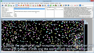
This tutorial video demonstrates how to perform automated cell counting
in images recorded in microplates showing a nuclear stain. This
particular recording was used for normalizing Seahorse cell respirometry data to cell counts.
Key features:
- Inhomogeneous background related to debris or tiling pattern is removed
- No constraint on maximal number of cells
- Results are directly transferred to Excel
Run time: 3 min 36 s.
Pipeline
 Unbiased and absolute calibration of ΔψM in millivolts
This tutorial video demonstrates how to work out millivolt values
of the mitochondrial and plasma membrane potentials in intact
adherent cells. Automated image and data analysis of
recordings using TMRM and the FLIPR membrane potential assay kit*
(aka PMPI) is demonstrated using pipelines and the Membrane
Potential Calibration Wizard. The example data relates to our recent
publication (26),
and the image files are available for download
here. See corresponding protocol
here.
Key features:
- The menu-activated pipeline performs all required image processing: background subtraction, image registration, spectral unmixing and automatic ROI drawing.
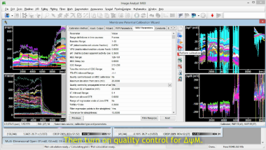
- Basic usage of the Membrane Potential Calibration Wizard allows point-and-click entering the frame ranges of calibrant additions, and converts each fluorescence trace into a millivolt trace.
- Advanced usage of the Membrane Potential Calibration Wizard allows setting up quality control based on estimated error of calibration.
Run time: 11 min 40 s.
*The FLIPR membrane potential assay kit is from Molecular Devices.
Protocol


